Parathyroid

Anatomy & Function

Central Neck Dissection

Parathyroid Anatomy & Function

Bean-shaped, rice grain sized gland, weighing 30 – 40 mg.

Vast majority of people are born with 4 parathyroid glands.

~ 1% have five or more glands (supernumerary).

~1% have < 4 glands.

They are located on the posterior surface (back side) of the thyroid gland, two on each side (lobe) of the thyroid.

One superior (higher along the thyroid, closer to the jaw) and one inferior (lower along the thyroid, closer to the collar bone) gland on each side.

  • Due to variability in location with regards to height in the neck, a more reliable relationship is relative to the recurrent laryngeal nerve (controls the movement of the vocal cord – contributing to voice, breathing, and swallowing) based on embryologic development.
    • Superior glands are always deep (closer to the spine) to the recurrent laryngeal nerve.
    • Inferior glands are always superficial (nearer to the skin) to the recurrent laryngeal nerve.

Parathyroid Anatomy

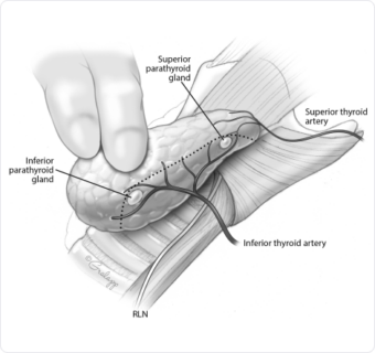
Image Source: Mary Ann Liebert, Inc.

Ectopic parathyroid glands (unexpected location) occur in 15-20% of patients.

The parathyroid glands originate high in the neck, then migrate lower down the neck during development as a fetus.

During this migration, some parathyroid glands may over-descend (located lower than anticipated) or under-descend (located higher than anticipated).

More common in inferior glands given the longer path of descent and traveling along with the thymus which can drag the inferior parathyroid glands with it lower into the upper chest (mediastinum).

Rests of parathyroid cells

Small, loosely organized groups of parathyroid cells which are functional.

Most commonly located in the thymus (mediastinum).

Important when considering surgical options for Kidney Related Hyperparathyroidism.

Two different cell types:

Parathyroid chief cells – produce and store parathyroid hormone (PTH).

Oxyphil cells – thought to be support cells, true function is unknown.

To control calcium levels in the body (calcium homeostasis).

Calcium is important for keeping bones and teeth strong, allowing muscles to contract, keeping your heart beating in a normal rhythm, and helping your brain send messages throughout the body via the nervous system, as well as many other vital functions.

Because of its essential nature, calcium levels need to be kept in a tight range, otherwise dysfunction throughout the body can occur if levels are too high or too low.

‘Teeter – totter effect’ (inverse relationship) of Calcium and Parathyroid Hormone (PTH).

All four parathyroid glands are constantly measuring calcium levels in the blood.

  • Measured by the Calcium Sensing Receptor (CASR) protein, located on the surface of the parathyroid cells.

When the calcium levels drop below a certain level (‘set point’ – varies slightly for all individuals), the parathyroid glands recognize this.

  • In response, the parathyroid gland will produce and release PTH into the blood stream.
  • PTH increases calcium levels in the blood by:
    • Increasing calcium reabsorption in the kidneys (not allowing it to leak out into the urine).
    • Resorption (release) of calcium from the bones into the blood.
    • Increases activity of enzymes in the kidney to convert Vitamin D 25 (inactive) to Vitamin D 1,25 (active form).
      • The active form of Vit D 1,25 increases calcium absorption from the intestines into the blood.

When the parathyroid glands function appropriately:

  • Low calcium levels will stimulate increased PTH production and higher PTH levels in the blood.
  • High calcium levels will inhibit PTH production and result in lower PTH levels in the blood.
    • This ‘teeter – totter’ effect is continuously working to keep calcium levels within the normal range.

Schedule your consultation today

For Personalized and Expert Surgical Treatment
of Your Thyroid and Parathyroid